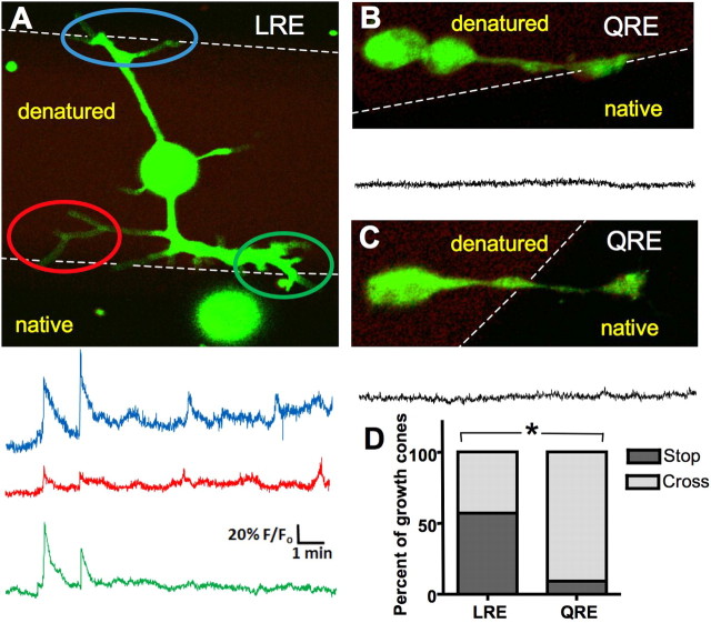
Figure 5.
Neurons at a border of laminin β2 generate calcium transients and stall. A, Neurons were cultured on stripes of native and UV-denatured laminin β2 LRE fragment and internal calcium concentrations were imaged using Fluo-4. Images were captured at 2 Hz. Fluo-4 is in green, denatured laminin β2 is in red, and native laminin β2 is in black. Three images were averaged to enhance visualization of the neuron. Traces show the calcium activity in each of three growth cones as they encounter the border. Axis scales apply throughout the figure. B, C, Less calcium transient activity is seen when a neuron approaches (B) or has crossed onto (C) activated point mutated laminin β2 (LRE→QRE). D, Significantly more neurons stop or turn back at an LRE border than a QRE border (*p < 0.001).

