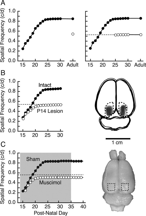
Figure 1.
Visual cortex-dependent enhancement of rat OKT from eye opening. A, Left, OKT SF threshold was low but measurable on P15 (filled circles) and by P20 increased to near the level of experimentally naive animals (open circle) (Douglas et al., 2005). Thresholds continued to increase until plateauing near P24–P25 and remained stable with repeated daily testing up to P30 and intermittent testing into adulthood. Right, Results of testing from P15 (filled circles) effectively replicated those in the left panel, whereas the thresholds of littermates with testing from P25 (open circles) did not differ from naive adults. The dotted line in this and other panels demarcates the OKT SF threshold of experimentally naive adult animals (open circle) in the left panel. B, Left, Bilateral aspiration of visual cortex on P14 (open circles; P14 Lesion) blocked the characteristic progression of OKT enhancement present in intact animals tested from P15 (filled circles; Intact). Right, Reconstruction of lesions in the P14 Lesion group from the left panel. Surface features of the brains and boundaries of lesions for each animal were traced in Adobe Illustrator, the area of individual lesions was filled with light gray, and the images were superimposed. Fiducial landmarks were used to estimate the borders of striate cortex (dashed lines) with reference to stereotaxic coordinates. C, Muscimol–Elvax treatment of visual cortex (period of implantation indicated with shading) blocked enhancement (open circles; Muscimol), resulting in a profile similar to that in animals with P14 lesions (B, left). Sham Elvax surgery did not affect enhancement (filled circles; Sham). Removal of Elvax had no effect on either group. Right, Surface view of a brain from a representative muscimol-treated animal in the left panel. The Elvax (approximate position demarcated with dashed box) caused no apparent tissue trauma (for additional examples, see supplemental Fig. 2, available at www.jneurosci.org as supplemental material).

