Figure 4.
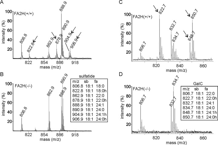
MALDI-TOF mass spectrometry. A–D Alkaline stable lipids isolated from the brain of FA2H+/+ (A, C) and FA2H−/− (B, D) mice were subjected to MALDI-TOF mass spectrometry in negative (A, B) or positive (C, D) ion mode, to detect sulfatide (A, B) and GalC (C, D), respectively. Mass to charge ratios (m/z) of individual sulfatide and GalC species are shown in the insets. Hydroxylated sulfatide and GalC species were not detectable in FA2H−/− mice. Mass peaks that correspond to hydroxylated lipids and are missing in FA2H−/− mice are indicated by arrows in A and C. sb, Sphingosine base; fa, fatty acid; h, hydroxylated. Hydroxylated sphingolipids were undetectable in samples of FA2H−/− mice.

