Figure 4.

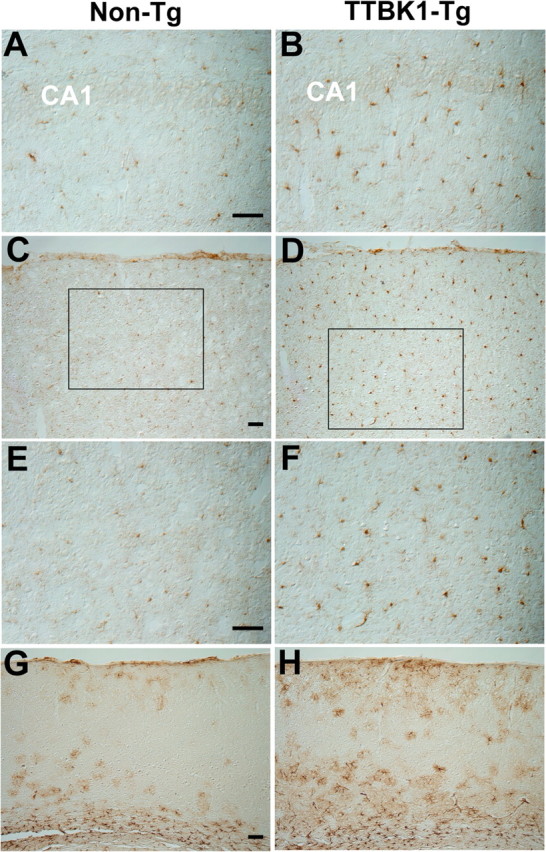
Microgliosis and astrogliosis in hippocampus and cortex of TTBK1-Tg mice. A–F, IBA1 staining of sagittal brain sections of aged non-Tg (A, C, E) or TTBK1-Tg mice (B, D, F) at 12–13 months old. A, B, hippocampus; C–F, visual cortex; E, F, high-power magnification of inset in C and D, respectively. G, H, GFAP staining of cortical region in non-Tg (G) and TTBK1-Tg mice (H). Original magnifications: (A, B, E, F), 200×; (C, D, G, H), 100×. Scale bars: (A, C, E, G), 100 μm.
