Figure 5.

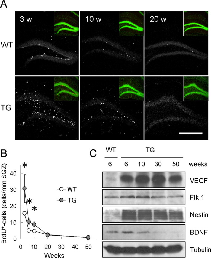
Reduced cell proliferation in older VEGF120-TG mice. A, Age-dependent reduction of cell proliferation in dentate gyrus. Animals at 3, 10, and 20 weeks (w) of age were killed 2 h after a single injection of BrdU. Proliferating cells in dentate gyrus of WT (top) and TG (bottom) mice were visualized by immunostaining for BrdU (white dots). Insets show nuclear staining (green) of the presented images. Scale bar, 500 μm. B, Quantification of proliferating cells along SGZ. The graph indicates the densities of BrdU-positive cells along SGZ at different time points (3, 6, 10, 20, and 50 weeks of age). Cell proliferation in dentate gyrus was reduced with age in both WT and TG mice. Cell proliferation in TG mice declined to the control levels by 20 weeks of age. *p < 0.05 by statistical analysis (see data). Data for BrdU-positive cell densities are as follows: 3 weeks old: WT mice, 15.50 ± 1.43 cells/mm SGZ; TG mice, 30.65 ± 8.02 cells/mm SGZ; n = 9 mice per group; p < 0.05, Mann–Whitney U test; 6 weeks old: WT mice, 4.98 ± 0.58 cells/mm SGZ; TG mice, 10.47 ± 1.63 cells/mm SGZ; n = 10 mice per group; p < 0.01, Welch's t test; 10 weeks old: WT mice, 4.74 ± 0.46 cells/mm SGZ; TG mice, 8.70 ± 1.48 cells/mm SGZ; n = 7 mice per group; p < 0.05, Welch's t test; 20 weeks old: WT mice, 2.51 ± 0.20 cells/mm SGZ; TG mice, 2.24 ± 0.81 cells/mm SGZ; n = 6 mice per groups; p = 0.11, Mann–Whitney U test; 50 weeks old: WT mice, 0.68 ± 0.19 cells/mm SGZ; TG mice, 0.96 ± 0.34 cells/mm SGZ; n = 4 mice per group; p = 0.50, unpaired Student's t test; independent experiments at each time point. C, Persistent expression of VEGF120 and Flk-1 (VEGF receptor 2) in VEGF120-TG mice. Hippocampal protein extracts from TG mice (6, 10, 30, and 50 weeks old) and WT mice (6 weeks old) were analyzed by Western blot for VEGF, Flk-1, Nestin, BDNF, and β-tubulin. The result showed consistent expression of VEGF120 (mature form is shown) and Flk-1 in TG mice. In contrast, the level of Nestin and BDNF tended to decline with age (see also supplemental Fig. 4, available at www.jneurosci.org as supplemental material, for quantitative analyses). β-Tubulin was used as an internal control.
