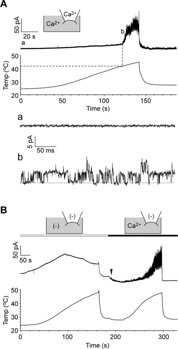
Figure 5.

Single-channel activation of Painless during heating. A, The top trace shows activation currents in Painless-expressing excised membrane during slow heating (0.2°C/s) in an inside-out patch-clamp mode at +60 mV holding potential (n = 4). The dotted line indicates an initiation point of the currents. Standard pipette solution (Ca2+ in the pipette) and Cs-Asp/200 nm Ca2+ bath solution (Ca2+ in gray area) were used. The basal trace in a and single-channel currents in b are magnified from corresponding lines in the top trace. The dotted line in b indicates the closed-channel level. B, Painless is robustly activated by heat in the presence of Ca2+ on the cytoplasmic side (n = 6). Ca2+(−) pipette solution [(−) in the pipette] and Cs-Asp/Ca2+(−) bath solution [(−) in gray area] or Cs-Asp/200 nm Ca2+ bath solution (Ca2+ in gray area) were used. Note that movements in basal lines including leak always occurred in the absence of Ca2+o and Ca2+i, but those are clearly different from single-channel currents observed in the presence of Ca2+i. Moreover, small but apparent single-channel currents were evoked as soon as Ca2+ was applied to the cytoplasmic side before heat application (arrowhead).
