
Figure 3.
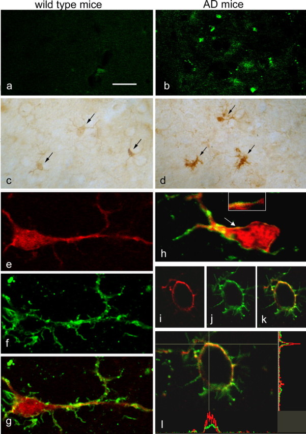
CLIC1 is expressed in microglial cells in wild-type aged mice and in the microglial cell membrane in an AD mouse model. a, b, At 18 months of age, thioflavin S staining reveals amyloid deposits (green spots) only in the brain of 3×TgAD mice (b). c, d, Immunoperoxidase labeling shows a more intense CLIC1 staining in cells morphologically identifiable as microglia (arrows) in AD mice (d) compared with wild type controls (c). e–l, Immunofluorescence double labeling shows that CLIC1 (e, h, i, l; red) is expressed in cells identified as microglia by their binding of LEA (f, h, j, l; green); g, h, k, l, Merged images. CLIC1 labeling is diffuse in ramified resting microglia from control mice (e–g; 9 positive cells of 75 analyzed; n = 3 mice), whereas in AD mice (h–l), it is present also in the cell membrane of activated microglia (arrow and inset in h) and of round phagocytic microglia (I–l; 65 positive cells of 84 analyzed; n = 3 mice). l, The enlargement of the cell shown in k is accompanied by intensity profiles along the horizontal and vertical lines demonstrating enrichment of CLIC1 labeling in the membrane stained by LEA. Scale bars: a–d, 25 μm; e–k, 10 μm; l, 8 μm.