
Figure 10.
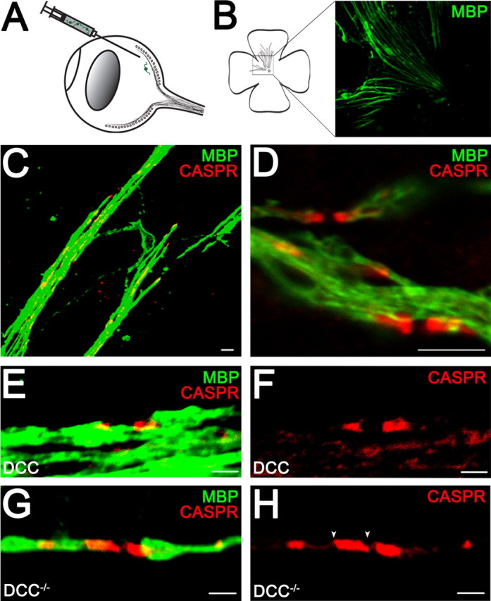
Altered paranodal Caspr distribution in retinal ganglion cell axons myelinated by DCC−/− oligodendrocytes in vivo. Injection of OPCs into the eyes of adult mice results in myelination (A). Flat-mounted retina (B) was double-labeled with antibodies against MBP to visualize myelin and Caspr to visualize paranodes (C–H). MBP was visualized using Alexa 488-conjugated antibodies (green). Caspr was visualized using Alexa 546-conjugated antibodies (red). The Caspr-immunoreactive domains were lengthened in the DCC−/− myelin group (G, H) compared with oligodendrocytes expressing DCC (E, F). The white arrowheads (H) illustrate the edges of the Caspr-immunoreactive domain measured. Magnification: B, 10× objective; C, 100× objective; D, 100× objective; digital zoom, 2; E–H, 100× objective; digital zoom, 4. Scale bars: C, D, 5 μm; E–H, 2 μm.