
Figure 5.
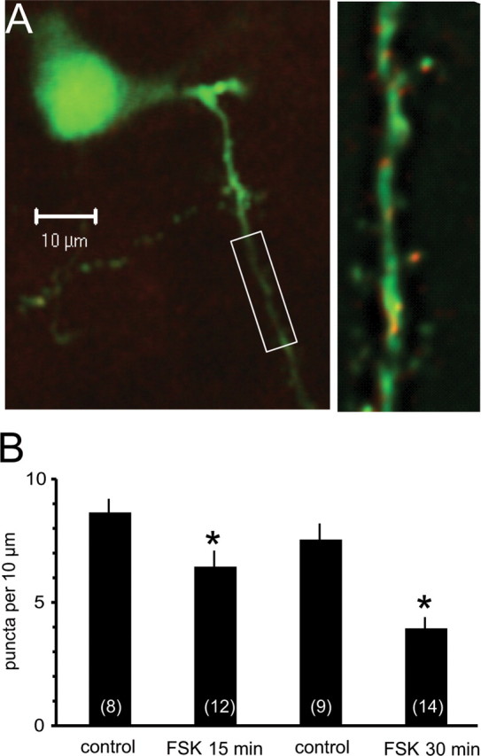
Activation of protein kinase A reduces surface expression of SK channels. Organotypic coronal brain slices were prepared and transfected using a gene gun. A, Principal neuron transfected with SK2 channels with primary and secondary dendrites as shown by venus fluorescence (green). SK2 channels visualized using anti-myc are shown in red and appear as puncta on the dendritic tree. The boxed section of the secondary dendrite is shown on a slightly enlarged view in the right panel. SK2 channel puncta were counted on primary and secondary dendrites and quantified as number of quanta per 10 μm of dendrite. B, Histogram shows mean data for the control group and after treatment with forskolin (FSK) for either 15 min or 30 min. Numbers of cells are shown in the bars. *p < 0.05.