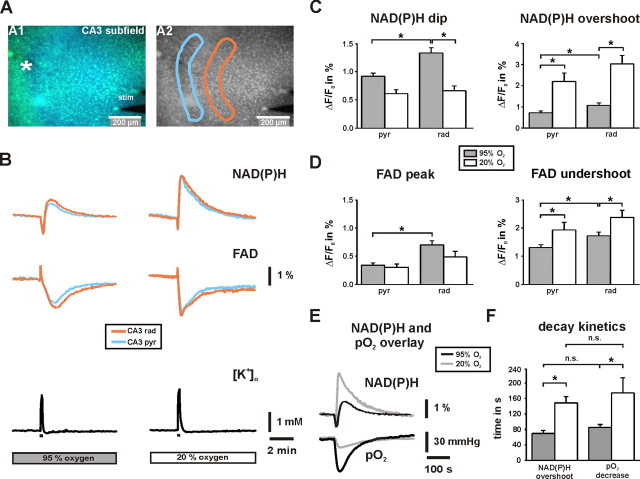
Figure 5.
NAD(P)H and FAD fluorescence transients in stratum pyramidale and stratum radiatum in CA3. A, Overlay of NAD(P)H and FAD fluorescence images as recorded with a delay of 130 ms. The K+-sensitive electrode was positioned in stratum pyramidale (white asterisk), the bipolar stimulation electrode close to the dentate gyrus (stim) for electrical activation of fiber tracts to CA3. Regions of interests (light blue, stratum pyramidale; orange, stratum radiatum) were selected to determine changes in %ΔF/F0 from image stacks that were recorded at 0.5 Hz. B, The shapes of biphasic NAD(P)H (top traces) and FAD (middle traces) fluorescence transients as evoked by stimulus trains (10 s, 20 Hz; black bars) are clearly distinct at 20% O2. Note that FAD transients (peak and undershoot) are inverse to NAD(P)H transients (dip and overshoot) because of different fluorescence properties of the dinucleotides. Transient increases in [K+]o as evoked by stimulus trains were simultaneously recorded (bottom traces) and indicate virtually the same degree of neuronal activation at 95 and 20% O2 (2.1 ± 0.1 and 2.4 ± 0.2 mm; n = 18; p = 0.26). C, D, Histograms summarizing the analysis of NAD(P)H and FAD transients (n = 18 each) in stratum pyramidale (pyr) and stratum radiatum (rad). Note that differences are most prominent in stratum radiatum. E, Overlay of NAD(P)H and pO2 transients in stratum pyramidale as evoked by stimulus trains. F, Histogram summarizing the decay time constants of NAD(P)H overshoots and pO2 transients at 95 and 20% O2 (n = 18). *p < 0.05.

