
Figure 10.
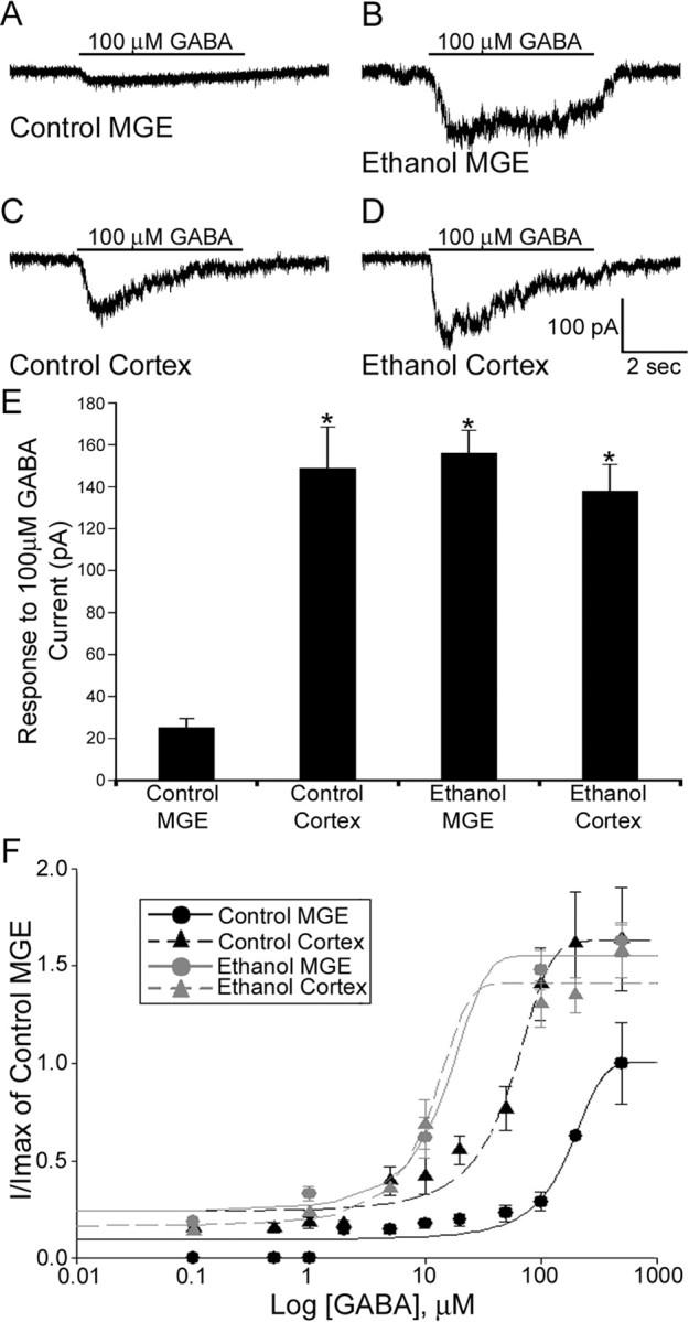
GFP+/MGE cells recorded in slices obtained from BAC-Lhx6 in utero ethanol-exposed embryos are more sensitive to GABA than those recorded within control slices. A–D, Responses to application of 100 μm GABA to GFP+/MGE cells recorded from the MGE (A, B) and neocortex (C, D) of control and in utero ethanol exposed acute BAC-Lhx6 slices, respectively. E, Average response of GFP+/MGE cells recorded within the MGE and the IZ of the neocortex in slices from control and in utero ethanol-exposed E14.5 embryos (control MGE, 25.25 ± 4.16 pA; control cortex, 148.85 ± 19.66 pA; ethanol MGE, 156.20 ± 10.72 pA; ethanol cortex, 138.06 ± 12.64 pA; ANOVA, p < 0.001). Data are expressed as mean amplitude ± SEM. Asterisks denote a significant difference from control MGE (p < 0.001). F, GABA concentration–response curves for GFP+/MGE cells recorded in the MGE and IZ of the neocortex of acute slices obtained from control and in utero ethanol-treated E14.5 BAC-Lhx6 embryos normalized to the max amplitude of cells recorded in the MGE of control slices.