
Figure 3.
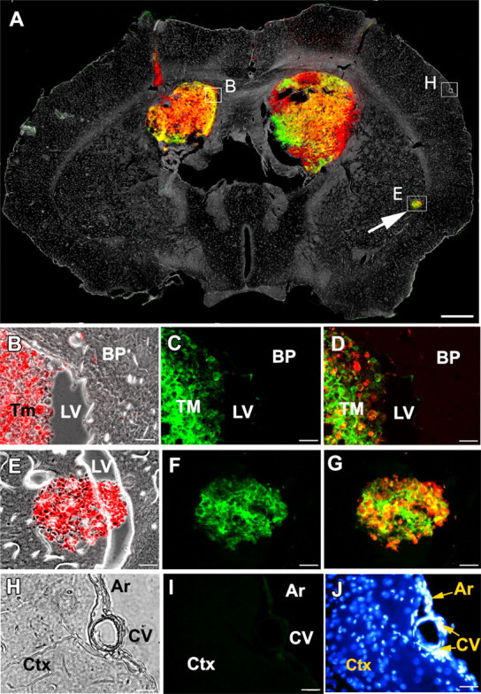
VSVrp30a targets multiple separate xenografts in the brain. A single intravenous injection of VSVrp30a was given to an SCID animal bearing bilateral striatal red fluorescent rU87 grafts and a third incidental tumor mass that has seeded through the CSF to the lateral ventricle. A is a montage of fluorescent and phase-contrast images from a representative coronal brain slice prepared 48 HPI. A shows colocalization of viral-encoded GFP and tumor cell-expressed RFP, simultaneously in all tumor masses. B–D were taken from the ventricular border of the right xenograft as marked on A. E–G are from the incidental tumor mass at the ventricular wall (arrow). H–J are from brain parenchyma, distant from the tumor graft. H shows phase-contrast image, and I shows the absence of viral fluorescence. J shows healthy nuclei of cells in the brain parenchyma (BP), arachnoid (Ar), and cortical blood-vessels (CV). Similarly, no infection was observed outside the tumor in the slice preparation at 72 HPI. Ctx, Cortex; LV, lateral ventricle; Tm, TM, tumor. Scale bars: A, 500 μm; B–J, 20 μm.