Figure 4.

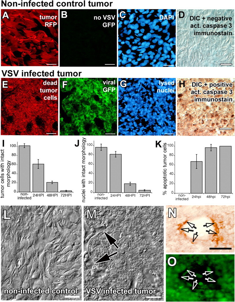
VSV infection of brain tumor xenografts results in widespread tumor cell death. Slice preparations from uninfected (A–D) and infected (E–H) rU87 xenografts in SCID mice. Uninfected tumor xenografts have normal cellular morphology as outlined by cytoplasmic RFP transgene expression (A), have no viral green fluorescence (B), have normal appearing nuclei (C), and do not stain with the apoptosis marker cleaved caspase 3 (D). VSV-infected tumor cells, in contrast, express viral GFP (F), lose cellular integrity (E), undergo chromatolysis (G), and stain positively for the apoptotic marker (H) at 48 HPI. Graphs (I–K) indicate the increase in viral tumor cell killing during the first 3 d after virus injection. Laser confocal DIC imaging shows uninfected (L) and infected (M) glioma xenografts. Note that, despite widespread cellular debris inside the VSV-infected tumor xenograft, vessel architecture is spared and endothelial cells appear morphologically healthy (arrows). Noninfected cells constituting the vessel wall stained positive for endothelial marker von Willebrand factor (arrows in N, O). Scale bar, 50 μm.
