
Figure 6.
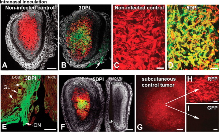
Gliomas within the olfactory bulb are infected after intranasal inoculation. Intranasally given VSV can enter the brain through the olfactory nerve. When rU87 glioma cells were unilaterally implanted into the olfactory bulbs of SCID mice, tumor xenografts grew similar to the striatal xenografts (A, C). Four days after implantation, animals were challenged with intranasal VSVrp30a and the virus was documented to infect the olfactory xenografts (B, D, F). VSV replication was documented in axons within the olfactory nerve (E, and arrow in B) and in some but not all of the olfactory glomeruli, which indicates the port of VSV entry (B, E). Virus can enter both tumor-containing and control bulbs through both olfactory nerves, but only tumor-bearing side shows infection in the central part (F). Simultaneously grown subcutaneous tumor grafts (G) did not show infection after intranasal VSVrp30a application (I), indicating that virus travels through the olfactory route rather than systemically. Stereomicroscopic picture of an intact subcutaneous flank tumor (G) and section of the same tumor (H). L-OB, Left olfactory bulb; R-OB, right olfactory bulb; EPL, external plexiform layer; GL, glomeruli; Mi, mitral cell layer; ON, olfactory nerve. Scale bars: A, B, G, 300 μm; E, F, 200 μm; C, D, H, I, 50 μm.