Figure 8.

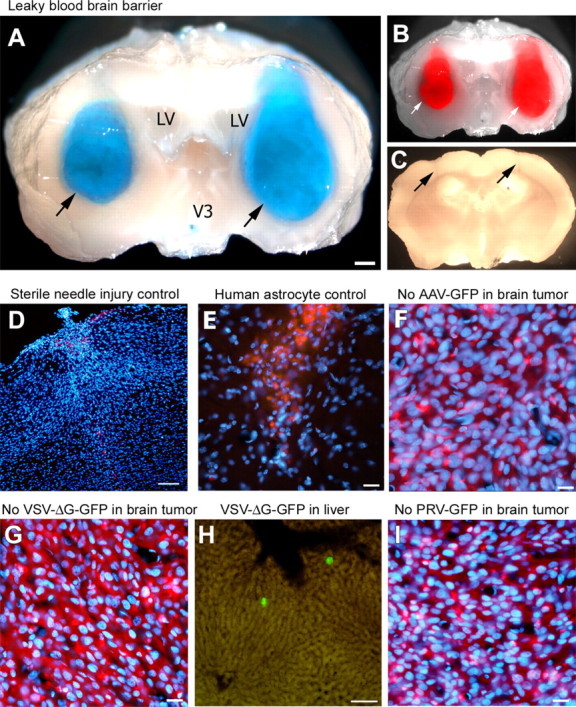
VSV selectively enters brain tumors through a leaky blood–brain barrier but does not infect control wounds or normal brain cell implants. After intravenous injection, Evans Blue leaked into tumor xenografts in which it colocalized (arrows) with tumoral RFP fluorescence, indicating an incompetent blood–brain barrier within the entire tumor (A, B). No Evans Blue leakage was observed in areas with needle injury only (see arrows) (C). Nonspecific brain damage inflicted by a sterile needle injury (D) or human astrocyte transplants (E) in the same brain region used for tumor grafting was not enough to allow VSVrp30a entry after intravenous injection of the same dose of VSVrp30a as in experimental animals. Tumor targeting in the brain required replication-competent VSV, and replication-deficient VSV (VSVdeltaG-GFP) did not infect rU87 xenografts in the mouse brain, but numerous infected cells were documented in the liver (G, H). No infection in the brain tumor, liver, or spleen was detected after intravenous injection of pseudorabies virus (PRV–GFP) or adeno-associated virus (AAV–GFP), both of which are known to infect glioma cells (F, I). LV, Lateral ventricle. Scale bars: A, 300 μm; D, H, 100 μm; E, G, 50 μm; F, I, 20 μm.
