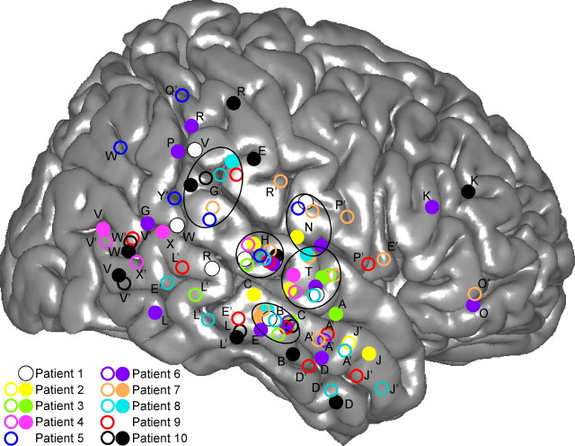
Figure 1.
Location of multicontact electrodes in the 10 patients, reported on a common 3D representation of the right hemisphere. The 3D representation of the cortex has been segmented from the anatomical MRI of the right hemisphere of the standard MNI brain. Open and filled circles are electrodes implanted in the left and right hemispheres, respectively. X and Y coordinates of each electrode of each patient have been normalized to the MNI space using the Talairach method. Letters correspond to the name prefix of electrode contacts. The names of left-implanted electrodes are followed by a prime.

