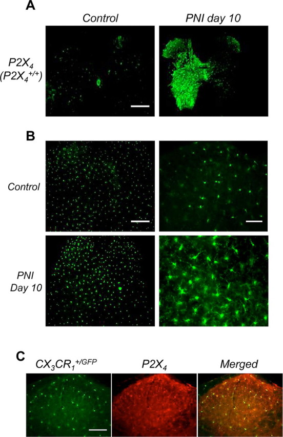
Figure 1.

Peripheral nerve injury induces upregulation of P2X4R in activated spinal microglia. A, Comparison of P2X4 expression in the spinal cord in sham-operated and injured mice. P2X4 immunoreactivity was barely detectable in sham animals whereas 10 d PNI, it was clearly induced ipsilateral to the lesion. Note that P2X4 expression spread to the whole side of the spinal cord because of sensory and motor fiber lesions. Scale bar, 500 μm. B, Morphology of spinal microglia in CX3CR1+/GFP mice post-PNI. Compared with sham animals, 10 d post-PNI microglia displayed higher fluorescence intensities, whereas the number of GFP cells was not significantly different. Increase of fluorescence intensity was restricted to the ipsilateral side of the lesion. Scale bar, 500 μm. At higher magnification in the dorsal horn region of the spinal cord (right), post-PNI microglia presented the typical morphological characteristics of the activated state with larger cell bodies and compacted processes compared with microglia from sham animals. Scale bar, 50 μm. C, PNI induces P2X4 expression in activated microglia. In the dorsal horn, P2X4 immunoreactivity colocalized exclusively with microglia-specific eGFP fluorescence in CX3CR1+/GFP mice 10 d post-PNI. Scale bar, 100 μm.
