
Figure 1.
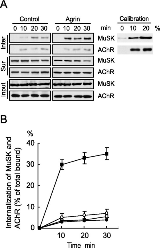
Agrin induces rapid internalization of MuSK. A, MuSK endocytosis in C2C12 cells in response to agrin stimulation. Cells were incubated with sulfo-NHS-SS-biotin to biotinylate surface proteins and subsequently challenged without or with agrin (5 nm) for indicated times. Cells were then incubated with the glutathione buffer to cleave biotin that remained on the cell surface. Internalized biotinylated proteins (Inter; top panels) were purified with streptavidin beads and analyzed by immunoblotting with anti-MuSK and anti-AChR antibodies. To monitor surface proteins, C2C12 cells were stimulated without or with agrin and incubated with sulfo-NHS-SS-biotin. Biotinylated surface proteins (Sur; middle panels) were purified with streptavidin beads and analyzed by immunoblotting. Bottom panels (Input) show equal amounts of MuSK and AChR in cell lysates as loading control. B, Quantitative analysis of data in A. Scanned autoradiograms were analyzed by the NIH Image software. The amounts of internalized proteins were calibrated with a standard curve of total biotinylated proteins (inset, right panels in A). Data shown were means ± SEM of three independent experiments. Squares, MuSK; circles, AChR (open, control; closed, stimulated).