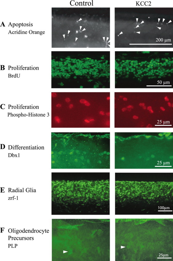
Figure 6.

Effects of KCC2 overexpression on apoptosis proliferation and differentiation in the zebrafish spinal cord. A, There is no obvious change in the levels of neurodegeneration during KCC2 overexpression because acridine orange-labeled apoptotic cells are not more abundant in the spinal cord of KCC2 embryos. Proliferation was not drastically modified by treatment because the number of BrdU-labeled cells (B) and the number of phospho-histone-3-labeled cells (C) is not significantly different between KCC2 and control embryos. In contrast, (D) neural differentiation was compromised in KCC2 embryos because a subset of committed progenitors marked with Dbx1 was significantly fewer in the spinal cord of these embryos. E, Zrf-1 staining of radial glial cells in the spinal cord of control and KCC2 embryos (n = 26 and n = 14, respectively). F, Fluorescent oligodendrocyte precursors in the spinal cord of PLP–GFP transgenics in non-injected zebrafish and zebrafish overexpressing KCC2 (n = 9 and n = 9, respectively). The arrowheads point to such precursors, which were hard to distinguish at this stage of development in the spinal cord.
