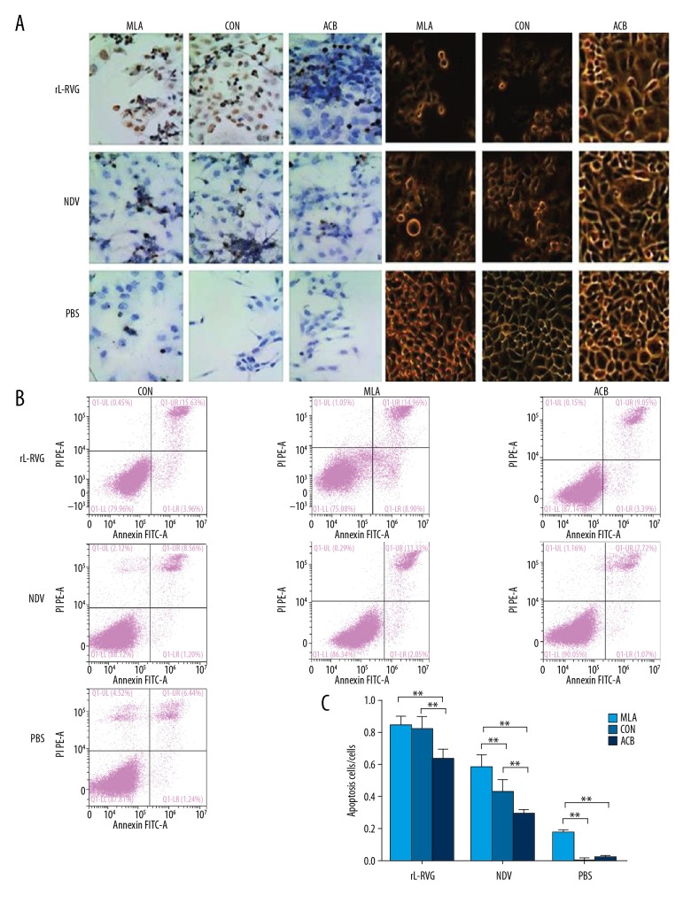
Figure 4.
Effect of rL-RVG, α7-nAChR antagonist, or agonist pretreatment on morphological changes and apoptosis in HGC cells. (A) Morphological changes of infected or uninfected HGC cells. When groups were treated by α7-nAChR antagonist MLA, the morphological changes in HGC cells were clearly more pronounced, including cell membrane shriveling and suspended growth, but the rL-RVG+MLA group showed little change. When groups were treated with α7-nAChR agonist ACB, the HGC cell morphological changes trended to persist, but there was no significant difference in morphological changes between the PBS+ACB group and the control group in comparison with the rL-RVG group. (B) TUNEL assay was used to assess apoptosis of infected HGC cells. (C) There were more apoptotic cells in the rL-RVG group than in the NDV group or PBS group (** P<0.01). When groups were treated with the α7-nAChR antagonist MLA, the apoptotic cells were more abundant than in the groups without pretreatment (P<0.01), except for the rL-RVG+MLA group and the rL-RVG group (P>0.05). When the α7-nAChR agonist ACB cells were pretreated before virus infection, the number of apoptotic cells decreased (** P<0.01), including early apoptotic (annexin V+/PI−) and late apoptotic (annexin V+/PI+) cells. * P<0.05.

