Figure 3.

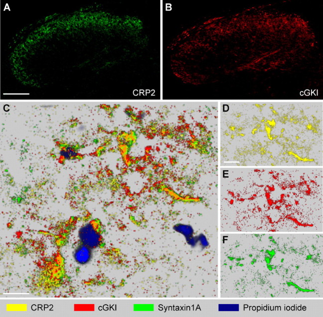
CRP2 and cGKI colocalization in the spinal cord. A, B, CRP2 and cGKI immunoreactivity in adjacent slices reveals a similar staining pattern of both proteins in the dorsal horn. C–F, Protein mapping by MELC in lamina II of the dorsal horn demonstrates colocalization of CRP2 (yellow) with cGKI (red) and syntaxin 1A (green). Extensive colocalization of CRP2 with cGKI appears orange. Propidium iodide (blue) was used to label DNA. Pictures were taken separately from the same slice using sequential rounds of fluorescent detection by the MELC robotic system and mapped using the software of the MELC system. C, Overview. D–F, Partition with single MELC maps of CRP2, cGKI, and syntaxin 1A. Scale bars: A (for A, B), 100 μm; C, D (for D–F), 10 μm. Maximal resolution in C–F was 0.4 μm (pixel size).
