
Figure 4.
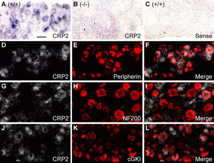
Expression of CRP2 in dorsal root ganglion cells. A–C, In situ hybridization for CRP2 mRNA (purple) in lumbar DRGs of wild-type mice (A) reveals CRP2 expression in a subset of DRG neurons. Specificity of the CRP2 mRNA antisense probe was confirmed by incubating DRGs of CRP2−/− mice with the antisense probe (B) and DRGs of wild-type mice with the respective sense probe (C). Pictures are presented as bright-field photomicrographs. D–L, Double labeling by a combined method of in situ hybridization for CRP2 mRNA and immunohistochemistry for peripherin (D–F), NF200 (G–I), or cGKI (J–L). CRP2 mRNA staining (D, G, J) was recorded as bright-field photomicrographs, converted to grayscale and inverted using Photoshop software. Thus, white indicates the presence of CRP2 mRNA. Immunoreactivity of peripherin (E), NF200 (H), and cGKI (K) was visualized by Cy3 fluorescence and appears in red. Overlays (F, I, L) demonstrate a large number of cells colabeling for CRP2 and peripherin (F) or cGKI (L) and a small number of cells colabeling for CRP2 and NF200 (I). Scale bar, 25 μm.