
Figure 2.
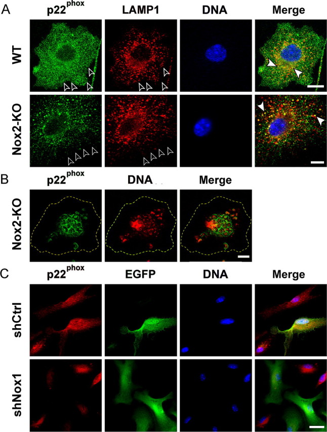
Nox1- and Nox2-dependent localization of p22phox in microglia. A, Codetection of p22phox (anti-p22phox, green), lysosomes (anti-LAMP1, red), and nuclei (Hoechst 33342, blue) in cultured WT or Nox2-KO microglia. Plasma membranes are outlined by open arrowheads. White arrowheads point to lysosomal p22phox in merged views (confocal microscopy). Scale bars, 10 μm. B, Detection of p22phox (green) and nucleic acid (Hoechst staining, blue fluorescence converted to red) in a Nox2-KO murine microglial cell after microglial phagocytosis of yeast particles (zymosan). The plasma membrane is outlined in yellow. Cultured cells were incubated with zymosan for 45 min before fixation and staining. Note the localization of p22phox in phagosome membranes surrounding stained nucleic acid of yeast particles (confocal microscopy). Scale bar, 10 μm. C, Suppression of p22phox expression in Nox2-KO microglial cells transduced with shNox1. Cultures of purified Nox2-KO microglia were transduced with lentiviral shNox1 or shCtrl and cultured for 3 d before fixation and codetection of p22phox (anti-p22phox, red) and EGFP (anti-EGFP, green). Cells transduced with lentiviral shRNA express EGFP. Scale bar, 20 μm.