
Figure 5.
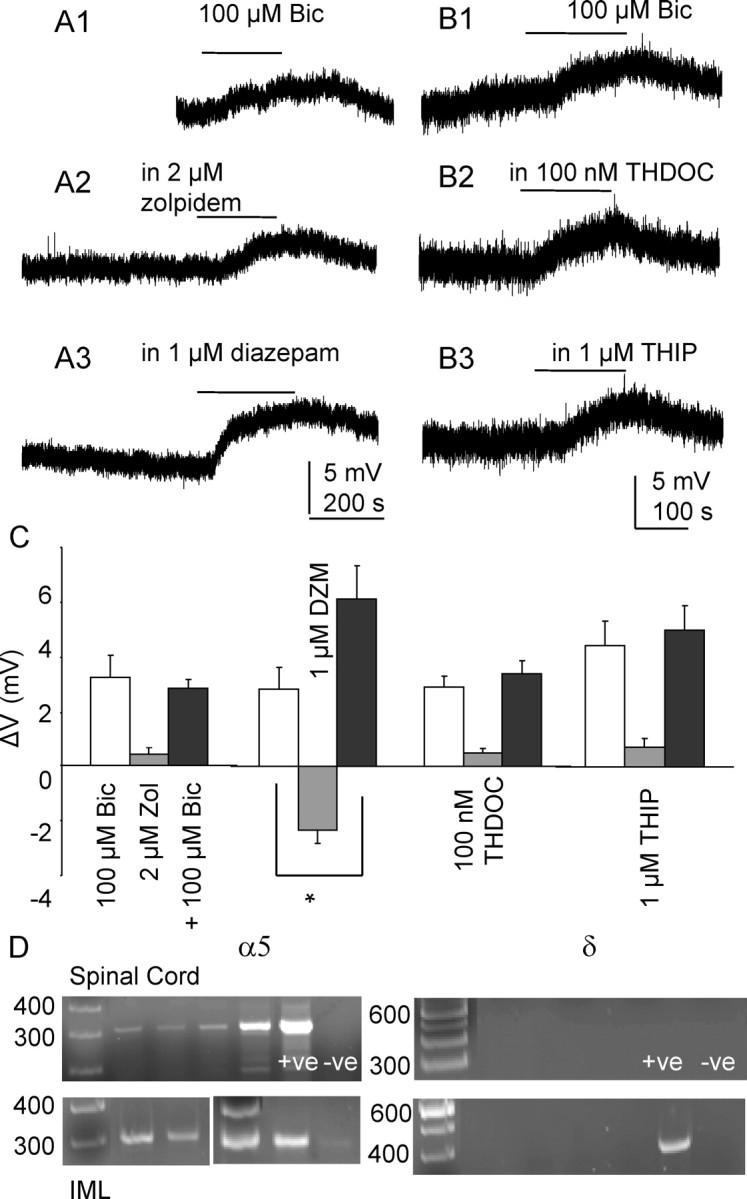
Tonic inhibition is mediated by α5 subunit-containing GABAA receptors. Recordings made in K-gluconate-based intracellular solution. Lines indicate application of 100 μm bicuculline. A1–3, Reponses to 100 μm bicuculline in the same neuron in control conditions, in 2 μm zolpidem and 1 μm diazepam (DZM). B1–3, Responses to 100 μm bicuculline in the same neuron in control conditions, in 100 nm THDOC and 1 μm THIP. C, Graph showing membrane potential changes to bath application of 100 μm bicuculline alone (white bars), modulators alone (gray bars), and bicuculline in the presence of a modulator (black bars). *p < 0.001, Student paired t test. Error bars represent SEM. D, Expression of α5 in the spinal cord and IML sample, denoted by the presence of a single band corresponding to the predicted size (300 bp). Amplicons representing the δ isoform are not detected in either the spinal cord or IML samples. This isoform is however present in the positive control tissue (+ve) taken from the hypothalamus, confirming the negative results observed are not due to the primers or amplification conditions.