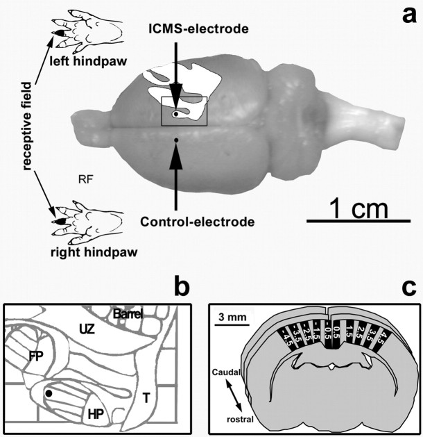
Figure 1.
Experimental procedure. a, The ratunculus with the hindpaw representation and the recording sites (dots) with a representative receptive field. An enlargement of the rectangle in illustration a is represented in b. It shows the place of the hindpaw representation in the map, which was electrically stimulated. c, For the investigation of the immunolabeled cells, at least nine coronal sections in a rostrocaudal extent of 2 mm with intervals of 250 μm between the sections were analyzed for each marker. Each coronal section was divided into sampling areas (five on each hemisphere) with a dorsoventral extent from the pia mater to the white matter and with a mediolateral extent of 500 μm in layer IV. HP, Hindpaw; UZ, unresponsive zone; FP, forepaw; Barrel, barrel cortex; T, tail.

