Figure 1.

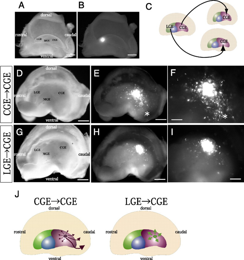
LGE cells behave differently from CGE cells when transplanted into the local environment of the CGE. A, B, Median view of the hemispheres. Focal electroporation of FITC-labeled oligonucleotides into the E13.5 LGE. Bright-field (A) and fluorescent (B) images of the telencephalic hemisphere indicated the site of electroporation and the specificity of labeling of the LGE. C, Schema of homotopic and heterotopic transplantations into the CGE in telencephalic hemisphere cultures. D–I, Bright-field (D, G) and fluorescent (E, F, H, I) images of the whole-mount hemispheres. Small fragments of the CGE (D–F) or LGE (G–I) taken from the locally electroporated E13.5 hemispheres and labeled with a CAG-driven DsRed-Express expression vector were transplanted into the CGE of other E13.5 hemispheres. The hemispheres were cultured for 40–42 h. F, I, Higher magnifications of E and H, respectively. While transplanted CGE cells migrated caudally toward the caudal end (asterisk in E, F), LGE-derived cells migrated only slightly in the CGE (H, I). A significantly smaller number of LGE-derived cells (14.8 ± 4.0% [SE], 4 independent experiments) than transplanted CGE cells (63.4 ± 7.9% [SE], 3 independent experiments) had migrated >500 μm (p = 0.0076). J, Summary of the experiments in which LGE cells or CGE cells were transplanted into the CGE. Scale bars: A, B, D, E, G, H, 500 μm; F, I, 200 μm.
