Figure 3.

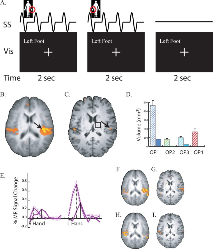
Responses to somatosensory stimulation. A, Stimulus and task paradigm. Three successive 2 s trials are shown. The somatosensory stimulus (row labeled “SS”) consisted of four repetitions of 200 Hz vibrotactile stimulation within a 250 ms ON/250 ms OFF square wave envelope delivered to the body part circled in red (left hand in first trial, right hand in second trial, no stimulation in third trial). The visual stimulus (labeled “Vis”) was identical in all trials. The behavioral task was to fixate the central crosshairs at all times, except when the left foot was stimulated, when the subject fixated the words “Left Foot.” B, An axial slice at the location of secondary somatosensory cortex in the operculum. Underlay shows average anatomical dataset from nine normal controls; colored voxels show significant responses to somatosensory stimulation (p < 0.01 corrected). Black arrow highlights strong opercular activity. C, Responses to somatosensory stimulation in the patient's operculum. Black arrow highlights weak activity. Black square shows location of right thalamic lesion. D, Volume of cortex responding to somatosensory stimulation in four cytoarchitectonic subdivisions of the operculum. The left hatched bar in each pair shows the mean ± SEM volume in nine normal controls; the right solid bar in each pair shows the patient. The label under each pair of bars identifies the subdivision. E, BOLD fMRI time course of the response to somatosensory stimulation. The black bar under the x-axis shows the duration of the 2 s stimulation trial for right hand stimulation (left trace) and left hand stimulation (right trace). Purple lines show the deconvolved event-related time series representing the average response to a single trial. Solid purple line shows the signal change in the peak operculum voxel of the patient (black arrow in C). Dashed purple line shows the peak signal change (mean ± SEM) in nine normal controls. Vertical black lines show the latency of the peak of the response (solid bar for patient, dashed bar for patient). F, Responses to left hand stimulation in the operculum of normal controls. G, Responses to left hand stimulation in the patient. H, Responses to right hand stimulation in the operculum of normal controls. I, Responses to right hand stimulation in the patient.
