
Figure 5.
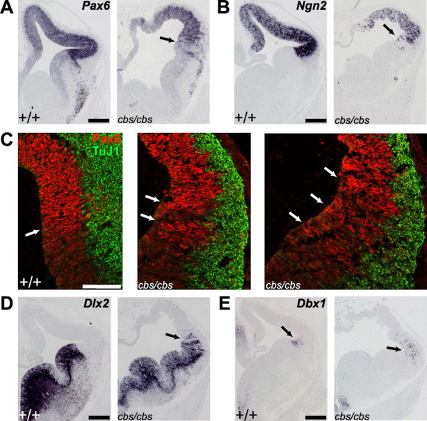
Relaxation of the pallial-subpallial boundary (PSPB) in cbs mutants. In situ hybridization (A, B, D, E) and immunohistofluorescence (C) analysis of 12.5 wild-type and cbs/cbs embryos. For each coronal section, one telencephalic half is shown, with dorsal to the top, lateral to the right. C, Red, Anti-Pax6 antibody; Green, TuJ1 antibody, recognizing newborn neurons. A, B, C, left panel, D, E, Arrows indicate the PSPB. C, Middle, right panels, Arrows indicate radial stripes of Pax6 expression at the PSPB in cbs/cbs mutants. A, Pax6. B, Ngn2. C, Pax6. D, Dlx2. E, Dbx1. Scale bars: A, B, D, E, 300 μm, (C) 100 μm.