
Figure 1.
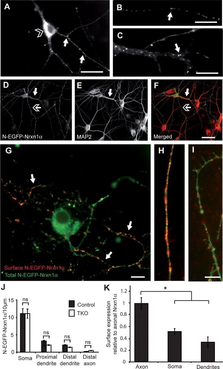
Epitope-tagged Nrxn1α occurs intracellularly and at the axonal surface of primary hippocampal neurons. A–C, N-EGFP-Nrxn1α is visualized by its autofluorescence, and is present in intracellular pools within the soma (open-headed arrow, A) and in puncta (white arrows) along axons (B) and dendrites (C). D–F, Colabeling of MAP2 (E, and red in F) to distinguish tagged Nrxn (D, and green in F) in dendrites (white arrow) and in axons (open-headed arrow). G–I, Colabeling of the surface population of EGFP-Nrxn1α (red). Surface Nrxn is mostly present at the plasma membrane of axons (H, G; white arrows) and less at soma (G) or dendrites (I). J, K, Quantification of total expression (J) and surface labeling (K) of Nrxn1α in different compartments (n = 4 transfected cultures); *p < 0.05. Scale bars: A, F, 40 μm; B, C, G, I, 20 μm.