
Figure 3.
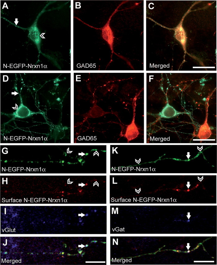
Excitatory and inhibitory neurons display the same pattern of Nrxn1α expression. A–F, Hippocampal neurons were transfected with N-EGFP-Nrxn1α on DIV7 and labeled with an antibody against GAD65 on DIV10 to distinguish between excitatory and inhibitory neurons. Both GAD65-positive (A–C) and GAD65-negative (D–F) cells show Nrxn1α expression in intracellular pools (open arrowheads) and as puncta along their processes (white arrows). G–N, Colabeling of surface Nrxn and the synaptic markers vGlut and vGat in N-EGFP-Nrxn1α-expressing neurons. Nrxn1α is transported along axons (G, K) and is inserted into the membrane (white arrows) as illustrated by extracellular GFP staining (H, L). Open arrowheads point to sites where Nrxn1α has not been inserted into the membrane, and is therefore present in transport vesicles. Membrane insertion occurs at excitatory and inhibitory synapses, as demonstrated by colocalization with vGlut (I) and vGat (M). Scale bars: A–F, 20 μm; G–N, 10 μm.