
Figure 4.
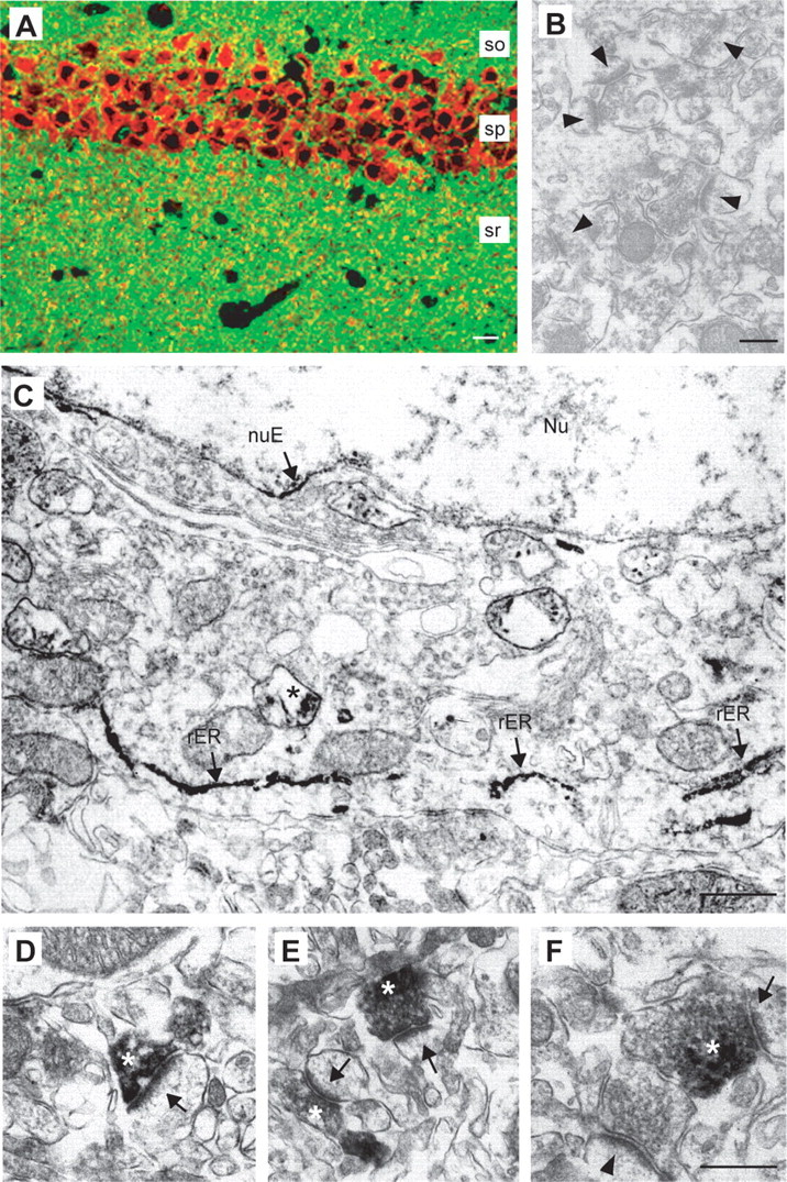
HRP-tagged Nrxn1α expressed in transgenic mice reveals polarized targeting mostly to presynaptic terminals. A, Double labeling of a hippocampal section with antibodies against the HRP-tag (red) and synaptophysin (green) shows strong expression of the transgene in pyramidal neurons of the CA1 region and partial colabeling of punctate clusters in the stratum radiatum (sr). so, Stratum oriens; sp, stratum pyramidale. B, C, Preembedding immuno-EM from nontransgenic (B, control, arrowheads point to unlabeled synapses) and Nrxn1α-HRP transgenic mice (C). C, Staining in somata is present in the rough endoplasmic reticulum (rER, arrows) and in vesicular structures (asterisk). nuE, Nuclear envelope; Nu, nucleus. D–F, Electron micrographs from the neuropil (sr of CA1) of transgenic mice show Nrxn-HRP at synapses (arrows), extending over the plasma membrane and part of the presynaptic terminals (asterisks). Scale bars: A, 40 μm; B, 500 nm; C, 800 nm; D, F, 250 nm (for D–F).