
Figure 6.
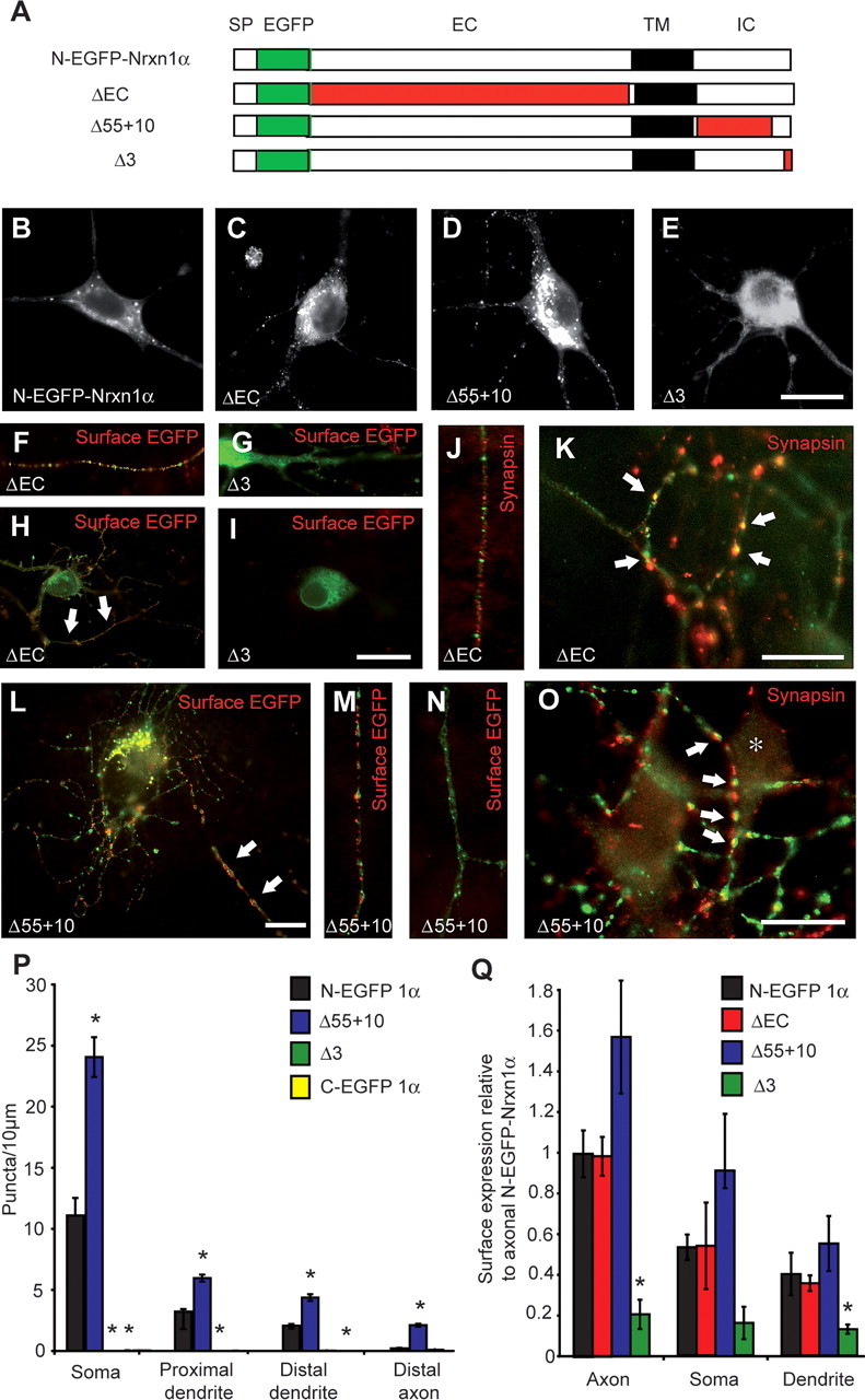
C-terminal sequences are involved in targeting of Nrxns. A, Schematic representation of full-length N-terminally EGFP-tagged Nrxn1α and of N-EGFP-Nrxn1α carrying various deletions (in red). SP, Signal peptide; EGFP, enhanced GFP tag; EC, extracellular region; TM, transmembrane region; IC, intracellular region. B–O, Hippocampal neurons were transfected with full-length or deletion constructs, and visualized by autofluorescence (B–E) or counterstained (in red) for surface EGFP (F–I, L–N) and synapsin (J, K, O). F, H, L, M, Nrxn constructs are inserted into the plasma membrane mainly in the axon (white arrows) except when the PDZ-binding motif was deleted (G, I). ΔEC and Δ55 + 10 constructs do not colocalize with synapsin in intracellular transport vesicles along the axon (J) but do so where appropriate synaptic contact sites are formed (K, O; white arrows). P, Quantification of Nrxn1α-positive puncta. Transfection of control (SKO) and triple (TKO) knock-out neurons with N-EGFP-Nrxn1α yielded similar levels and distribution. In contrast, the construct Δ55 + 10 was present at levels approximately twofold to threefold those of N-EGFP-Nrxn1α, and addition of a C-terminal tag (C-EGFP-Nrxn1α) or deleting the PDZ motif (Δ3) significantly reduced the number of puncta present at all subcellular locations (n ≥9 cultures). Q, Quantification of the surface expression of extracellularly tagged Nrxn1α constructs (relative to axonal N-EGFP-Nrxn1α) demonstrated that deleting the extracellular portion of Nrxn1α (ΔEC) had no effect on its surface presentation, whereas the construct Δ55 + 10 had increased surface labeling, and Δ3 was significantly decreased (n ≥ 3 cultures). *Significantly different to N-EGFP-Nrxn1α (p < 0.05). Scale bars, 20 μm.