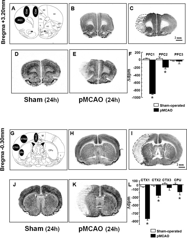
Figure 2.
Radioactive in situ hybridization of NCKX2 transcript in the cerebral cortex and caudate–putamen of sham-operated and ischemic animals 24 h after sham surgery or pMCAO. A schematic diagram of regions of interest at coronal bregma levels of +3.20 mm and of −0.30 mm are shown in A and G. Representative brain NeuN-immunohistochemistry-processed sections deriving from sham-operated and pMCAO-bearing rats are shown in B and C (bregma level, +3.20 mm) and H and I (bregma level, −0.30 mm), respectively. D and J and E and K represent the in situ hybridization autoradiographic film images obtained from sham-operated and ischemic brain sections with NCKX2 antisense probe, respectively. mRNA levels expressed as Δdpm ± SEM and presented for each region of interest analyzed, with white columns for sham-operated rats and black columns for pMCAO rats, are shown in F and L. Δdpm indicates the difference between the dpm value of each region of interest ipsilateral to the ischemic side and that of the contralateral corresponding side. *p < 0.05 versus respective values in sham-operated groups. aca, Anterior commissure, anterior part; AID, agranular insular cortex, dorsal part; AIV, agranular insular cortex, ventral part; Cc, corpus callosum; Cg, cingulate cortex; CPu, caudate–putamen; DP, dorsal peduncular cortex; DTT, dorsal tenia tecta; fmi, forceps minor of the corpus callosum; IL, infralimbic cortex; I, insular cortex; LO, lateral orbital cortex; M, motor cortex; M1, primary motor cortex; M2, secondary motor cortex; Pir, piriform cortex; PrL, prelimbic cortex; VO, ventral orbital cortex; VTT, ventral tenia tecta. Scale bars: C (for B, C), I (for H, I), 2 mm.

