
Figure 8.
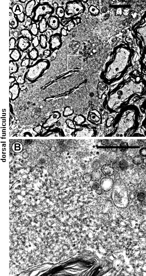
Electron microscopic features of spheroids. A, B, Dorsal funiculus of a iPLA2β−/− mouse at 56 weeks. B shows a higher magnification of the boxed region in A. Tubulovesicular structures can be seen in the spheroid, as well as degenerated vesicles and vacuoles. Scale bars: A, 2 μm; B, 0.5 μm.