
Figure 4.
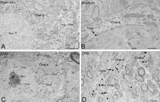
Ultrastructural localization of CHOP/GADD153 immunoreactivity in hypoglossal neurons. A–D, EM photomicrograph reveals CHOP/GADD153 silver-labeled particles (arrow) were rarely evident in ChAT-labeled somata (A; ChAT-S) or dendrites (B; ChAT-d) in sham LTIH mice. In contrast, exposure to LTIH (8 weeks) was associated with increased CHOP/GADD153-labeled particles within nuclei and throughout the soma and dendrites of ChAT peroxidase-labeled motoneurons (C, arrows). Within the nucleus (nuc) and nucleolus, CHOP/GADD153 colocalizes within subregions of increased electron density. Within dendrites of mice exposed to LTIH, CHOP/GADD immunoreactivity increased significantly. Scale bars: A, C, 2 mm; B, D, 0.5 mm.