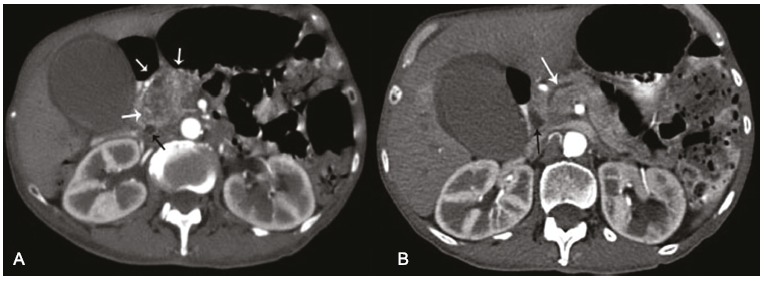
Figure 1. – Axial computed tomography of the abdomen in the arterial phase. A - Heterogeneous mass in the topography of the head of the pancreas (white arrows) and dilation of the common bile duct (black arrow); B - Dilation of the pancreatic duct (white arrow) and the common bile duct (black arrow).

