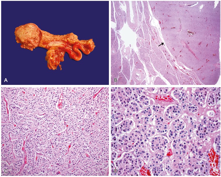
Figure 3. – A - Gross macroscopic view of the somatostatinoma located in the head of the pancreas - histological appearance of the pancreatic somatostatinoma; B - Panoramic photomicrography of the tumor showing the well circumscribed border (arrow) and the normal pancreatic parenchyma (HE – 25×); C - Photomicrography of the tumor cells arranged in a trabecular or ribbon pattern, separated by delicate fibrovascular stroma (HE – 100×); D - Photomicrography of tumor cells with a relatively uniform and round shape, with fine granular eosinophilic cytoplasm and a central located round nucleus (HE – 400×).