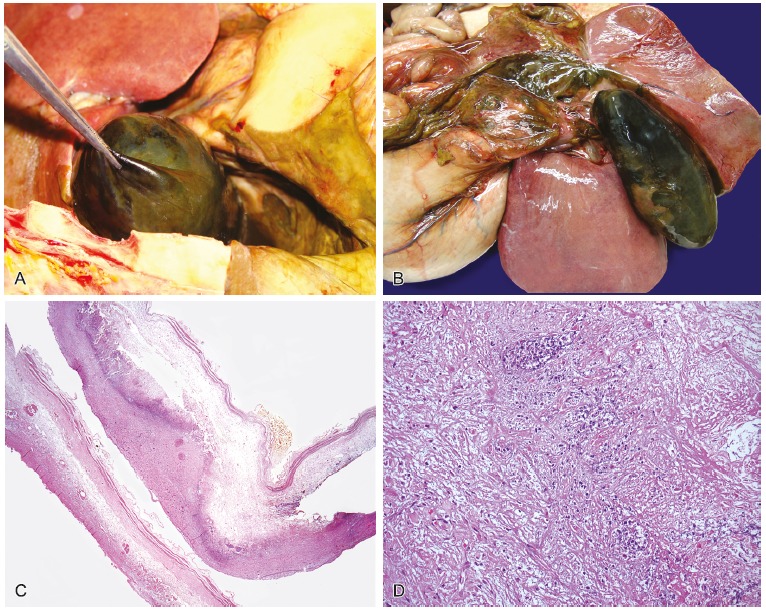
Figure 5. – A and B - Gross macroscopic view of distended gallbladder with dark greenish and external surfaces, with deposition of friable material, surrounded by a thick collection; C - Photomicrography of the gallbladder showing evident autolysis and acute inflammatory infiltrate (HE – 25×); D - Photomicrography of the gallbladder wall showing, in detail, the acute inflammatory infiltrate (HE – 400×).

