
Figure 1.
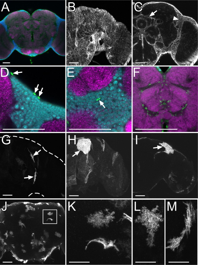
Repo-positive glial cells in the adult central brain. A, Glial nuclei labeled by anti-Repo antibody (green), neuronal nuclei labeled by anti-Elav (blue), and neuropil labeled by nc82 (magenta). B, C, Glial membrane labeled with mCD8::GFP driven by repo-GAL4 on brain surface (B) and inner brain (C). Arrow and arrowhead show glial membrane forming borders among substructures of neuropil and brain cortex, respectively. D–F, Glial nuclei (green) on brain surface (D), cortex (E), and neuropil (F). Glial nuclei (arrows in D and E), neuronal nuclei, and neuropil were labeled as shown (A). G–I, Single cells of small surface glia (G), large surface glia (H), and cortex glia (I) labeled by FLP-out combined with repo-GAL4. J–M, Single cells of neuropil glia labeled by FLP-out combined with repo-GAL4. A dendritic and fibrous lamellar morphology of neuropil glia was labeled around neuropil (J). Two different types of neuropil glia arranged nearby (square in J) are shown at a high magnification (K). L, M, High-magnification images of a dendritic (L) and fibrous lamellar morphology (M) of neuropil glia. Scale bars: A–J, 50 μm; K–M, 25 μm.