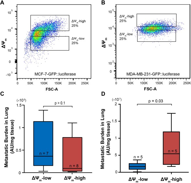
Figure 7.
Correlation of ΔΨm with metastatic potential in vivo. (A) MCF-7 cells and (B) MDA-MB-231 cells, both transduced with GFP/luciferase, were sorted into a ΔΨm-high and -low subpopulations for tail-vein injection into NSG mice; Quantification of the metastatic burden in the lungs of mice injected with (C) MCF-7 cells at week 5 and (D) MDA-MB-231 cells at week 4, via ex vivo quantification of luciferase activity in tissue lysate (p-values: Mann-Whitney test).

