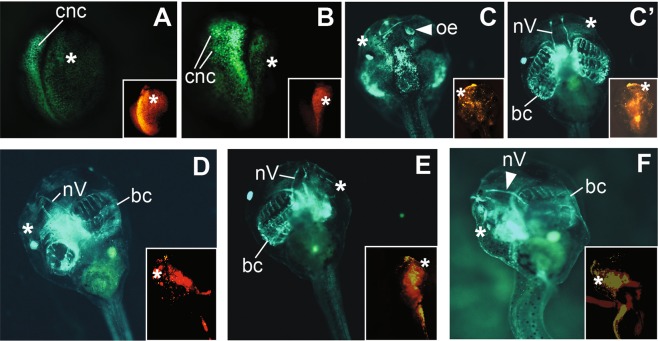
Figure 4.
Phenotypes of ADAM13 knockdown displayed by snai2:eGFP embryos. Eight-cell stage heterozygous snai2:eGFP embryos were injected with 1.5 ng MO 13-3 to target ADAM13 in one dorsal-animal blastomere, and cultured to the indicated stages; a red fluorescent dye was co-injected as a lineage tracer. The injected side is denoted with a white asterisk, and structures that are present on the uninjected side but absent on the injected side are denoted with white arrowheads. Insets show red fluorescence images of the same embryos. (A,B) Dorsal view (with anterior at the top) of stage ~18 (A) and ~22 (B) embryos displaying reduced CNC domain on the injected side, as determined by eGFP expression. In (B) CNC migration is normal on the uninjected side but inhibited on the injected side. (C–F) Injected embryos that did not show apparent defects in CNC induction or migration were selected and cultured to stage ~46. (C,C’) are dorsal and ventral views (with anterior at the top), respectively, of the same tadpole. The olfactory epithelium (oe) is not detectable (C) but branchial cartilage (bc) and trigeminal nerve (nV) appear normal (C’) on the injected side of this embryo. (D,E) Embryos with under-differentiated head cartilage structures on the injected side, as compared with the uninjected side. (F) An embryo with severely defective trigeminal nerve and head cartilage structures on the injected side. (D–F) are ventral views with anterior at the top.

