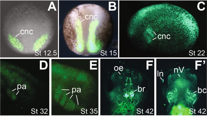
Figure 5.
Wnt signaling activity in the CNC lineage. Heterozygous transgenic Wnt reporter embryos were imaged at the indicated stages. Expression of eGFP is detectable in the pre-migratory (A,B), migrating (C) and differentiating (D–F’) CNC. (A,B) dorsal view with anterior at the top; green fluorescence and bright-field images are merged to show the relative positions of CNC in the whole embryo. (C–E) side view with anterior to the left (C) or right (D,E). (F,F’) dorsal and ventral views (with anterior at the top), respectively, of the same tadpole. br, brain; ln, lens; bc, branchial cartilage; nV, trigeminal nerve; oe, olfactory epithelium; pa, pharyngeal arches.

