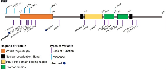
Figure 1.
Schematic 2D representation of the PHIP and its functional domains. Locations of likely pathogenic/pathogenic variants from previously reported individuals and the new variants reported herein indicated in bold. Schematic does not include splice site (three) or translocation (one) variants. (Adapted from Webster et al. 2016.)

