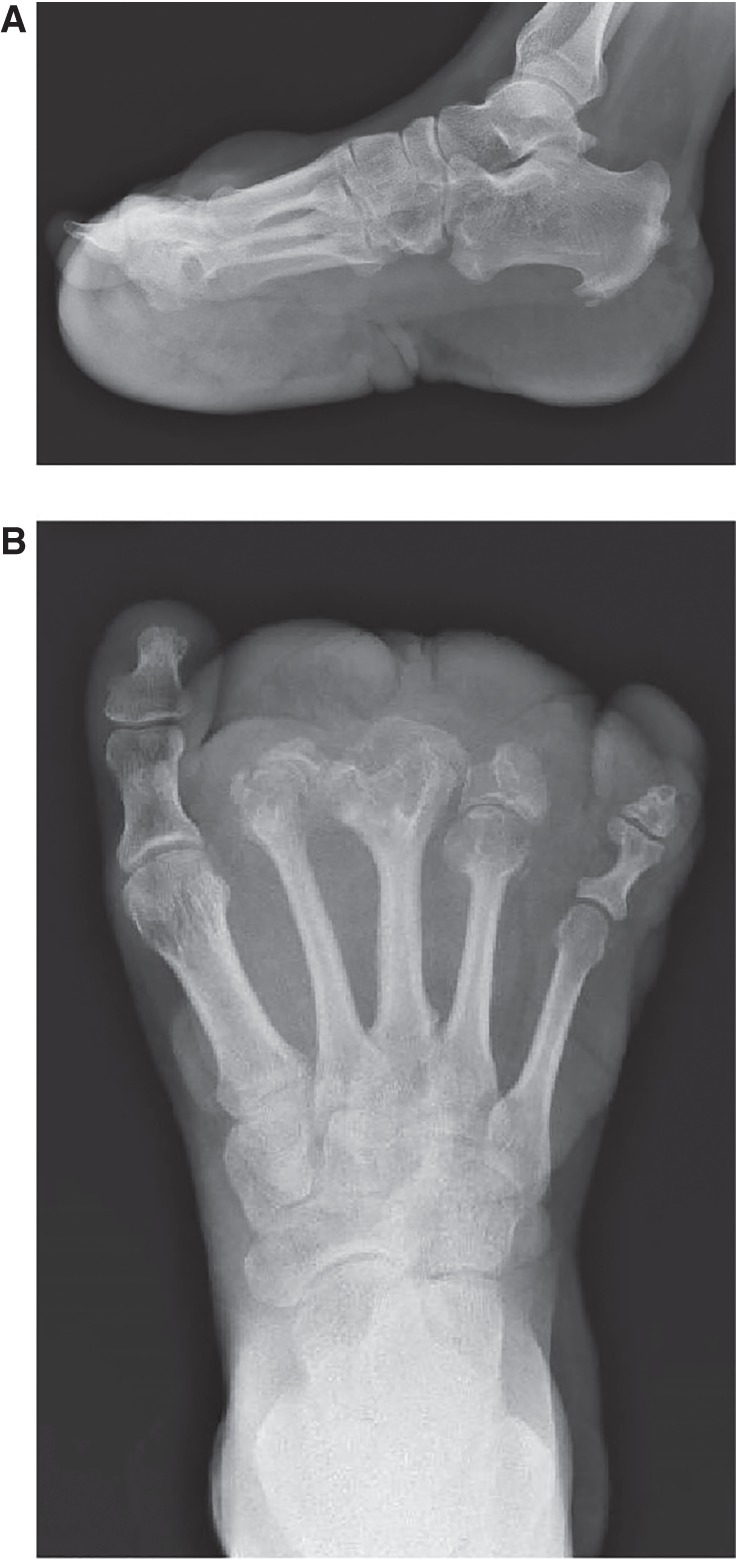
Figure 2.

Individual 1 (OG.194). (A) Lateral and (B) AP radiographs of her left foot demonstrated partially absent second, third, and fourth phalanges, widening of the second to fourth metatarsal heads and shafts, calcific enthesopathy of the insertion of Achilles’ tendon and plantar fascia, and prominence of plantar, midfoot dorsal soft tissue.
