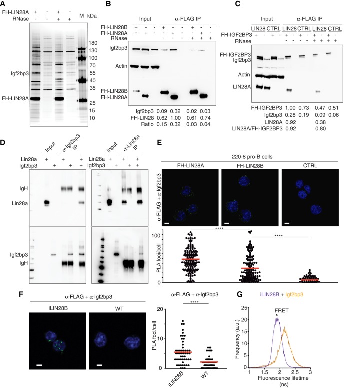
Figure 2.
Igf2bp3 and Lin28b proteins form an RNP complex. (A) Flag immunoprecipitation (Flag-IP) of lysates treated with or without RNase A from 220-8 pro-B cells transduced with FH-LIN28A (lanes 1,3) or mock-transduced 220-8 cells (lanes 2,4) and marker (M) fractionated by SDS-PAGE and silver-stained. Samples from the same immunoprecipitations were analyzed by mass spectrometry (Supplemental Table 3). (B) Flag-IP of lysates treated with or without RNase A from 220-8 cells either mock-transduced or transduced with FH-LIN28A or FH-LIN28B subjected to Western blotting and probed with anti-Igf2bp3, anti-Actin, and anti-HA antibodies. Background-corrected intensities for the indicated bands are listed below in arbitrary units along with ratios (Igf2bp3/FH-LIN28). Western blotting of corresponding amounts of input material from the same lysates serves as comparison. (C) Western blotting of input and Flag-IP material from lysates from 220-8 cells transduced first with either empty vector (MSCVpuro) or untagged LIN28A and then with either empty vector (MSCVneo) or FH-IGF2BP3 and probed with anti-Igf2bp3, anti-Actin, and anti-LIN28A antibodies. (D) Recombinant Lin28a and Igf2bp3 proteins purified from E. coli were coincubated in vitro, immunoprecipitated, and probed on a Western blot with the indicated antibodies. (E) The in situ proximity ligation assay (PLA) was performed on 220-8 cells transduced with either empty vector (WT), FH-LIN28A, or FH-LIN28B to detect Igf2bp3 proteins and Flag epitopes that are within ∼40 nm of each other. (Top row) Representative confocal microscopic images show fluorescent foci (green) that result from PLA. Nuclei were stained with DAPI (blue). Scale bar, 5 µm. (Bottom row) The number of fluorescent foci per cell is plotted for each sample. At least 90 cells from five random fields were counted per sample. (****) P ≤ 0.0001, Mann-Whitney U-test. (F) PLA was performed on adult BM pro-B cells from iLIN28B mice or WT littermate controls using anti-Flag and anti-Igf2bp3 primary antibodies similar to E. (****) P ≤ 0.0001, Mann-Whitney U-test. (G) Fluorescence lifetime in nanoseconds is plotted in a histogram in the presence of the donor only (anti-Igf2bp3 only; orange) or in the presence of a Förster resonance energy transfer (FRET) partner (anti-Igf2bp3 and anti-Flag costaining; purple) in adult BM pro-B cells from an iLIN28B mouse.

