
Figure 3.
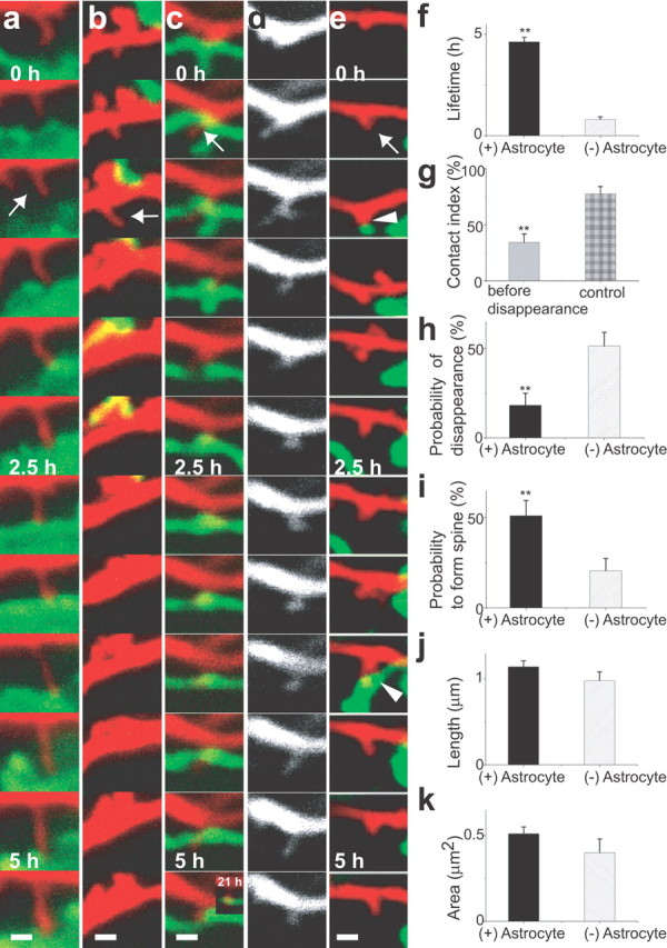
Enhanced lifetime and maturation of dendritic protrusions by astrocytic contacts. a–e, Fates of dendritic protrusions (red) with (a, c, e, arrows) or without (b, arrow) astrocytic contacts (green). Dendritic protrusions persisted with astrocytic contacts (a, c, e) but retracted without contacts (b). New dendritic protrusions (c, e) showed either prolonged (c) or transient (e, arrowheads) astrocytic contacts. In both cases, dendritic protrusions were subsequently stabilized. The same dendritic protrusion is presented with different colors in c (red) and d (white). f, Enhanced lifetime of dendritic protrusions with episodes of astrocytic contacts calculated from 5.5 h image sequences [n(DP) = 38/12 (with (+)/without (−) Astrocyte, for all data in this legend]. g, Reduced contacts with astrocytes in the 1 h period before retraction of dendritic protrusions [n(DP) = 38]. h, i, Astrocytic contacts reduce disappearance of dendritic protrusions [n(DP) = 33/30] and increase the probability to form spines [n(DP) = 32/30]. j, k, Length and area of dendritic protrusions with or without astrocytic contacts [n(DP) = 260/142]. All data are shown as mean ± SEM. **p < 0.01. Scale bars, 1 μm. DP, Dendritic protrusion.