
Figure 1.
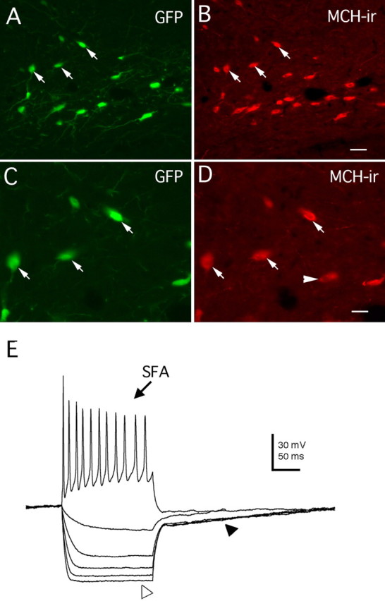
GFP expression restricted to MCH-containing neurons in transgenic mice. A, GFP expression in a hypothalamic section of a GFP–MCH transgenic mouse. Arrows indicate three cells. B, The same three cells (arrows) in A are shown, where antisera reveal immunoreactive MCH (MCH-ir; Texas Red) in the same neurons that express GFP, confirming that GFP expression in this line of transgenic animals is restricted to MCH-synthesizing neurons in the lateral hypothalamus. C, D. The same three cells shown in A and B are depicted at higher magnification. E, Intrinsic membrane properties of MCH neurons identified by GFP expression. SFA, Spike-frequency adaptation (after a 60 pA positive current injection for 200 ms). The open arrowhead indicates the inward rectification induced by −30 to −150 pA current injection for 200 ms, 30 pA steps; the filled arrowhead indicates the outward rectification observed in these hypothalamic cells. Scale bars: A, B, 20 μm; C, D, 10 μm.