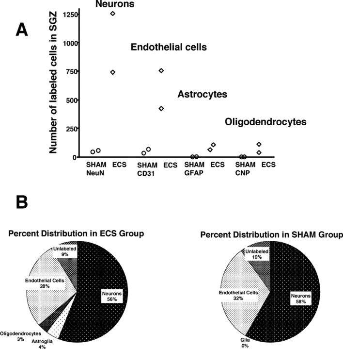
Figure 6.
Maturational fate of proliferating cells in the SGZ. A, B, The hippocampal sections of animals (2 ECS and 2 sham) killed 4 weeks after injection of BrdU were double-labeled for BrdU and maturational markers for mature neurons (NeuN), astroglia (GFAP), oligodendrocytes (CNP), or endothelial cells (CD31). Confocal analysis was conducted on a minimum of 25 BrdU-immunoreactive cells in at least three separate hippocampal sections to determine the percentage double labeled with each of the maturational markers. These percentages were then multiplied by the total number of BrdU cells per millimeters cubed of the SGZ that was previously determined using peroxidase methods. A, Graph showing the number of each mature cell-type for each animal. B, Pie charts showing the different percentages of mature cells in the ECS and sham groups.

