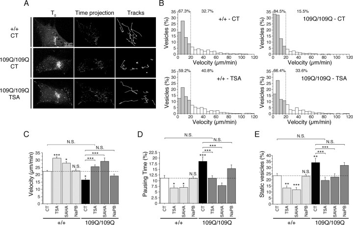
Figure 1.
TSA and SAHA stimulate vesicular transport of BDNF and rescue transport defect in HD Cells. A, B, WT (+/+) and 109Q/109Q cells transfected with BDNF-eGFP were treated for 4 h with DMSO (0.1%, CT), TSA (1 μm), or SAHA (2 μm) and analyzed by videomicroscopy. A, The displacement of BDNF vesicles is reduced in 109Q/109Q compared with +/+ cells as shown by 3D reconstruction of the first time point (T0), time projection of moving structures over the 1 min experiment, and visualization of 10 paths followed by individual vesicles (Tracks). The altered BDNF transport in 109Q/109Q cells (CT) is restored by treating cells with TSA. B, Analysis of the distribution of vesicular velocities shows a marked increase in the number of vesicles that have a low velocity in 109Q/109Q cells compared with +/+ cells. The distribution of velocities in TSA-treated 109Q/109Q cells is similar to the distribution in +/+ cells. Filled bars correspond to nonmoving and low-moving vesicles, and open bars correspond to high-speed moving vesicles. C–E, WT (+/+) and 109Q/109Q cells transfected with BDNF-eGFP were treated for 4 h with DMSO (0.1%, control, CT), TSA (1 μm), SAHA (2 μm), or NaPB (10 mm) and analyzed by videomicroscopy. Dynamics were quantified by the use of three parameters: the mean velocity of vesicles (C), their pausing time (D), and the percentage of static vesicles (E). Dashed lines correspond to the control values. N.S., Not significant. *p < 0.05; **p < 0.01; ***p < 0.001.

