
Figure 7.
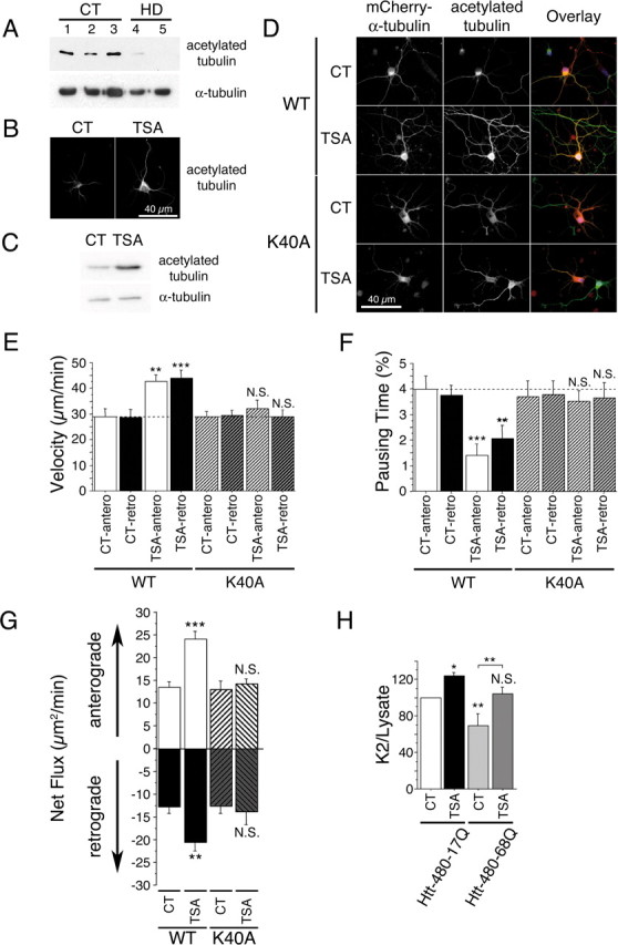
Tubulin acetylation at lysine 40 stimulates bidirectional transport and rescues BDNF release in HD. A, Acetylated tubulin levels are decreased in the brain of HD patients. Protein extracts are prepared from whole striatum of CT (samples 1–3) and HD individuals (HD grade 3, sample 4; HD grade 4, sample 5) and analyzed by immunoblotting for acetylated and total α-tubulin. B, C, Cortical neurons were treated with DMSO (0.1%, CT) or TSA (1 μm) for 4 h and analyzed by immunofluorescence (B) and immunoblotting (C) for the presence of acetylated tubulin. D, Cortical neurons were transfected with WT mCherry-α-tubulin or mCherry-α-tubulin K40A, treated for 4 h with DMSO (0.1%, CT) or TSA (1 μm), and analyzed by immunofluorescence for the presence of mCherry-α-tubulin (red) and acetylated tubulin (green). E, F, Cortical neurons were cotransfected with BDNF-eGFP and either WT mCherry-α-tubulin or mCherry-α-tubulin K40A, treated for 4 h with DMSO (0.1%, CT) or TSA (1 μm), and anterograde and retrograde vesicular movements in neurites were analyzed by videomicroscopy. Dotted lines correspond to the control values. G, TSA treatment increases anterograde and retrograde flux in a lysine 40-dependent manner. H, Cortical neurons were electroporated with BDNF and either htt-480–17Q or htt-480–68Q and treated for 4 h with DMSO (0.1%, CT) or TSA (1 μm). Transport-dependent BDNF release after a second KCl-induced depolarization is expressed as a K2/lysate ratio. N.S., Not significant. *p < 0.05; **p < 0.01; ***p < 0.001.